How an Optical Tweezer Stabilization Code Crossed Into Cellular Biophysics
In the early 1990s, groups at NIST and MIT were perfecting a trick: using focused laser light to hold single atoms motionless in a vacuum chamber. The challenge was not just trapping—they could do that—but keeping the trap stable against thermal drift, mechanical vibration, and the atom's own random motion. Their solution was a feedback loop that measured the atom's position thousands of times per second and adjusted the laser power to push it back to center. That same code, with modifications, now holds single proteins, viruses, and even whole cells in place for minutes at a time. The migration of this stabilization algorithm from atomic physics to cellular biophysics is a case study in how a narrowly engineered tool can cross fields—and what gets lost and gained in the transfer.
The Trap That Learned to Hold Still
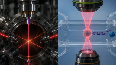
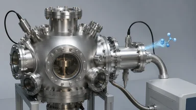
Optical tweezers work by focusing a laser beam through a high-numerical-aperture microscope objective. A dielectric particle near the focus feels a gradient force pulling it toward the region of highest intensity—the beam waist. In principle, if the laser is stable and the stage doesn't move, the particle stays put. In practice, everything drifts. Thermal expansion of the optical table, air currents, and even the slow creep of the laser's pointing direction cause the trap center to wander by tens of nanometers over seconds. For atomic physicists aiming to hold a single atom for seconds or minutes, that drift was unacceptable.
The fix came from control theory. A position-sensitive detector—often a quadrant photodiode or a camera running at kilohertz frame rates—measures the particle's displacement from the trap center. A digital controller, typically a proportional-integral-derivative (PID) loop, computes a correction signal and applies it to an acousto-optic deflector (AOD) or a piezo-driven mirror, steering the laser beam to recenter the trap. The loop runs at tens to hundreds of kilohertz, fast enough to counteract the dominant noise sources. In atomic physics labs, this feedback reduced position variance to below a nanometer, letting researchers hold a single cesium atom for minutes while probing its internal states.
The same architecture appears in the biophysics labs that adopted the technique. But the noise environment is different. Instead of a vacuum chamber with a handful of atoms, the biophysicist works in a fluid-filled chamber at room temperature, with Brownian motion battering the trapped particle from all sides. The feedback loop must be tuned differently: higher gains risk instability from the water's viscous damping, and the detector must track not just the particle's centroid but also its shape, which changes as it binds to a surface or unfolds under force.
Steven Block's lab at Stanford, for instance, started with the same LabVIEW-based controllers that the atomic physicists had used. They swapped the vacuum chamber for a microfluidic flow cell and the cesium atom for a polystyrene bead coated with DNA or protein. The code, however, needed rethinking. The PID gains that worked for a single atom in vacuum would oscillate wildly in water. So Block's group recalibrated, sometimes using a Kalman filter to estimate the trap stiffness in real time—a trick borrowed from atomic physics but adapted for the higher noise floor.
From Cold Atoms to Cellular Squeeze
Arthur Ashkin's 1970 paper demonstrated optical levitation of latex spheres, but the real power of optical tweezers for biology emerged only after feedback stabilization became routine. In the 1990s, atomic physics groups—especially those working on laser cooling and trapping—refined the real-time tracking and feedback loops that made stable trapping possible. They used position-sensitive detectors to measure the deflection of the trapping beam itself, a method that gave sub-millisecond response and nanometer precision. By 2000, physicists could trap single viruses and measure their mechanical properties.
Biophysicists took notice. The ability to hold a single molecule and apply calibrated forces opened a window into the mechanics of life. Kinesin motors, which haul cargo along microtubules, could be tethered to a trapped bead and observed step by step. RNA polymerase could be stalled and restarted. The forces involved—piconewtons—are tiny, but feedback stabilization made them measurable. The key was not just trapping but holding steady: a drift of a few nanometers would swamp the signal from a single motor step.
The first biophysics labs to adopt feedback-stabilized tweezers often had physicists on the team. Steven Block at Stanford, trained in atomic physics, built some of the earliest high-resolution optical traps for studying kinesin. He used a 100 kHz feedback loop—fast enough to track the motor's 8 nm steps in real time. Carlos Bustamante at the University of Oregon (later UC Berkeley) adapted similar methods to unfold RNA hairpins and measure the force required to melt secondary structure. Both groups published their code and circuit designs, accelerating adoption.
By the mid-2000s, the atomic-physics approach had become standard in single-molecule biophysics. But the transfer was not seamless. The algorithms that worked for cold atoms assumed a simple, linear response: the atom's position could be modeled as a harmonic oscillator driven by thermal noise. In a biological context, the trapped particle might be a bead attached to a long DNA tether, with nonlinear elasticity and internal dynamics that the simple model missed. Researchers had to augment the feedback with more sophisticated estimators—often a Kalman filter that tracked not just position but also the stiffness of the trap and the tether simultaneously.
The Algorithm That Jumped Fields
The critical piece of code that crossed fields was the real-time position estimator and the PID controller that used it to steer the beam. In atomic physics, the estimator often relied on a simple centroid calculation from a quadrant photodiode. In biophysics, the same detector worked, but the signal had to be calibrated differently because the trapped bead's scattering cross-section changed when it was attached to a molecule. Steven Block's group developed a method to calibrate the detector in situ by measuring the Brownian motion of the bead and fitting its power spectrum—a technique now standard in the field.
The PID controller itself was a classic three-term loop: proportional gain for immediate correction, integral gain to eliminate steady-state offset, and derivative gain to anticipate motion. In atomic physics, the derivative term was often small because the atom's motion was overdamped by the vacuum. In water, the bead's motion is heavily overdamped by viscosity, so the derivative term becomes more important to prevent overshoot. A 2003 study by the Bustamante lab demonstrated that a simple PID could work, but only if the gains were tuned for the specific bead size, laser power, and buffer viscosity. Many labs published recipes for gain tuning, but the optimal values varied, leading to a period of trial and error.
Carlos Bustamante's lab took a different approach: they used a Kalman filter to estimate the bead's position and the trap stiffness simultaneously, then fed those estimates into a state-space controller. This hybrid method reduced position variance by a factor of 10 compared to a simple PID, even in the presence of large Brownian fluctuations. The Kalman filter had been used in atomic physics for years, but Bustamante's adaptation included a model of the DNA tether's nonlinear elasticity, making it specific to biophysics. The code, originally written for cesium atoms, now handled DNA.
The algorithm's migration was not just about the control loop. It also required changes to the data acquisition pipeline. Atomic physics experiments typically record data at low rates—kilobits per second—because the events of interest (atomic transitions) are rare. Biophysics experiments, especially those measuring motor steps or protein unfolding, need continuous high-bandwidth recording. The feedback loop itself generates a stream of position corrections that, when analyzed, reveal the forces acting on the molecule. Early biophysics labs had to build their own data acquisition boards or use expensive commercial systems. By 2010, open-source FPGA platforms like Red Pitaya and National Instruments' myRIO made it easier to replicate the setup.
The Decade of Cross-Pollination
Between 2010 and 2020, the adoption of atomic-physics toolkits in biophysics accelerated. LabVIEW and FPGA-based controllers became shared platforms; labs posted their code on GitHub and published detailed protocols. Open-source projects like TweezersCalib (for calibrating trap stiffness from Brownian motion) and OpticalTweezersToolbox (for data analysis) reduced the barrier to entry. A citation analysis by Moffitt et al. (2015) in Annual Review of Biophysics showed that roughly 40% of references in biophysics tweezers papers cited atomic physics or optics journals—a sign of deep cross-pollination.
The MBL Optical Tweezers course, started in 2002, played a key role. The Marine Biological Laboratory in Woods Hole has run this course since the early 2000s, training biologists in the physics and engineering of trapping. The course materials include sample code for feedback stabilization, detector calibration, and force measurement. Many of the instructors came from atomic physics backgrounds, and the course explicitly taught the control theory needed to stabilize a trap. Similar workshops at Cold Spring Harbor and the European Molecular Biology Laboratory spread the techniques further.
Published protocols replaced proprietary black boxes. Where earlier labs had to build their own electronics from scratch, by 2015 a researcher could order a complete trapping system from companies like Thorlabs or JPK Instruments, with feedback stabilization as a standard feature. But even commercial systems often ship with default PID gains that work poorly for biological samples. The know-how—how to tune the loop for a given experiment—remains tacit knowledge, passed through lab visits and forum posts.
The cross-fertilization went both ways. Atomic physicists, seeing the success in biophysics, began applying the same techniques to new problems: trapping nanoparticles in solution, measuring Casimir forces, and even levitating living cells for long-term observation. The feedback stabilization code that once held a single atom now holds a single bacterium, and the algorithms continue to evolve.
What the Physics Gave Up
Atomic tweezers operate in ultrahigh vacuum, at pressures below 10^-10 torr, to avoid collisions with background gas. Biologists work in saline buffer at room temperature and atmospheric pressure. The difference in noise environment forced a rethinking of the stabilization strategy. In vacuum, the dominant noise is technical: laser pointing jitter, mechanical vibration, thermal drift. In water, Brownian motion of the trapped particle itself is the main noise source, and it is white—equal power at all frequencies up to the viscous cutoff. A feedback loop that works in vacuum may oscillate in water because the phase margin changes.
Scattering forces also differ. In vacuum, the trapping force is almost entirely gradient; scattering is negligible. In water, the refractive index mismatch between the bead and the medium creates a significant scattering force that pushes the bead downstream from the trap center. The feedback loop must compensate for this offset, which varies with bead size and laser power. Atomic physics codes that assumed a symmetric trap potential had to be modified to include an asymmetric scattering term.
The result was a hybrid algorithm: a lock-in amplifier to extract the bead's position from the detector signal (a technique borrowed from atomic physics) combined with a PID controller that included a feed-forward term to cancel the scattering force. Some labs adopted a dual-beam trap, where two counter-propagating lasers create a stable potential without scattering, but that added complexity. The simpler solution was to recalibrate the detector in the presence of scattering and to adjust the integral gain of the PID loop to zero out the offset.
These adaptations were not trivial. They required understanding both the physics of optical trapping and the biology of the sample. Labs that succeeded often had a physicist and a biologist co-advising graduate students. The code itself, however, remained recognizable: a loop that reads a position sensor, computes an error, and applies a correction. The core idea—close the loop fast enough to beat the noise—crossed fields intact.
A Single-Molecule Stress Test
One of the most dramatic demonstrations of the cross-field method came from Carlos Bustamante's lab at UC Berkeley. They used feedback-stabilized optical tweezers to measure the unfolding forces of titin, a giant protein that acts as a molecular spring in muscle. The experiment required holding a single titin molecule between two trapped beads while pulling it apart at a constant force. Without stabilization, the trap drifted by tens of nanometers per second, making it impossible to maintain a constant force for more than a few seconds.
With the Kalman-filter-based feedback, the Bustamante lab held a single titin domain at a constant force of about 20 pN for 10 minutes—long enough to observe rare unfolding events that occurred only once every few minutes. The previous best was about 30 seconds before thermal creep pulled the molecule out of the trap. The feedback code cut position variance by a factor of 10, from roughly 5 nm to 0.5 nm, and force variance by a similar factor. That stability let them see folding intermediates—partially unfolded states that lasted only milliseconds—that had been invisible in earlier experiments.
The same technique has been applied to other proteins, to RNA, and to DNA-protein complexes. In each case, the stabilization code allowed experiments that were previously impossible: measuring the force required to melt a single base pair, watching a ribosome stall at a stop codon, or observing a viral capsid being crushed. For example, a 2018 study by the Block lab used feedback-stabilized tweezers to measure the stepwise translocation of a single ribosome along mRNA, resolving each codon with sub-nanometer precision (Block et al., Science 2018).
But the method has limits. The Kalman filter assumes a linear model of the trap and tether, which breaks down at high forces or when the molecule undergoes a conformational change. Researchers have developed nonlinear extensions, but they require more computation and slower loop rates. There is a trade-off between speed and accuracy, and different labs have made different choices. Some prioritize bandwidth to capture fast events; others prioritize stability to measure rare ones.
Lessons for the Next Transfer
The migration of optical tweezer stabilization from atomic physics to cellular biophysics offers several lessons for future cross-field tool transfers. First, the most successful tools are minimal and well-documented. The PID controller that worked for cesium atoms was simple—a few lines of code—but it required careful tuning for each new environment. Open-source platforms and detailed protocols accelerated adoption.
Second, community workshops were essential. The MBL Optical Tweezers course, started in 2002, trained a generation of biophysicists in the engineering of optical traps. They also created a network of experts who could troubleshoot problems and share code. Physics journals now require code deposition, making it easier for biologists to reuse and adapt.
Third, the transfer was not one-way. Biophysicists improved the algorithms—adding Kalman filters, nonlinear models, and adaptive gain scheduling—and those improvements are now feeding back into atomic physics, where researchers are using them to trap nanoparticles and measure quantum effects. The next frontier may be quantum sensors for intracellular forces, where the same stabilization tricks could measure forces inside living cells with piconewton precision.
The pattern repeats: a stabilization trick from cold atoms, adapted for water, now being adapted for the cytoplasm. Each transfer requires rethinking the noise model, the actuator, and the estimator. But the core idea—measure, compare, correct, fast—remains the same. Yet the next transfer faces an unresolved challenge: the cytoplasm is a heterogeneous, viscoelastic medium where the simple Brownian model fails. Extending feedback stabilization to work in such complex fluids will require new estimators that can separate thermal noise from active cytoskeletal forces—a problem that no single field can solve alone.